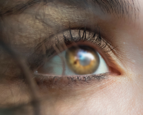
How an Eye Exam Can Identify Glaucoma
Did you know that a comprehensive eye exam catches glaucoma long before you develop symptoms? And if you wait until symptoms like vision loss appear, it’s too late because the optic nerve has already suffered permanent damage.
Routine eye exams at VIP Laser Center in Palm Beach Gardens, Florida, catch glaucoma at an early stage. As a result, you can start treatment that slows down or stops the gradual progression of glaucoma and prevents vision loss.
When to get an eye exam
All adults should have a baseline eye exam when they turn 40. After that, the frequency of your follow-up exams depends on your vision, eye health, and risk factors for glaucoma.
The risk factors for glaucoma include:
- Age over 60
- Diabetes
- High blood pressure
- Farsightedness
- Thin cornea
- Long-term use of corticosteroids
- Family history of glaucoma
- African, Hispanic, or Asian heritage
- Previous eye injury or surgery such as cataract surgery
The best way to determine how often you need an eye exam is to schedule an appointment and let us check your vision and screen your risk for glaucoma. Then we can recommend an eye exam schedule that protects your eye health.
Glaucoma eye exam
When performing a glaucoma eye exam, we include five essential tests:
Tonometry test
The tonometry test measures the pressure inside your eyes, called intraocular pressure (IOP). IOP is the most important risk factor for glaucoma. At least 90% of people with glaucoma have high IOP, a problem that occurs as fluids inside the eye build up.
As IOP increases, the pressure pushes against the optic nerve. Without treatment to restore normal eye pressure, more of the optic nerve becomes permanently damaged and you slowly lose your vision.
Dilated eye exam
After applying eyedrops to dilate the pupil, we use a small device that magnifies our view of the structures inside your eye. During this exam, we can see optic nerve damage and typically take an image of the nerve. This allows us to compare changes in the nerve at your next appointment.
Along with high pressure, visible damage to the optic nerve is the earliest sign of glaucoma.
Visual field test
Your visual field refers to how much you can see when you focus on a central point. In addition to testing your central vision, this test reveals any blind spots you may have and evaluates your peripheral (side) vision.
For most people, the loss of peripheral vision is the first noticeable symptom of glaucoma.
We routinely include visual field testing in your regular eye exam if you’re at risk of vision loss due to glaucoma. Monitoring changes in your visual field shows the severity and progression of glaucoma.
Gonioscopy
Gonioscopy is a simple test that shows the angle where your iris meets the cornea. The angle marks the spot where fluid inside your eye, called the aqueous humor, leaves your eye through a drainage system.
In a healthy eye, the fluid drains out at the same pace that new fluid enters the eye, maintaining a constant, normal pressure. Pressure builds up when too much fluid enters your eye and/or too little fluid drains out.
If the angle is open but you have high pressure, as it is with open-angle glaucoma (the most common type of glaucoma), the drainage system is blocked.
Pachymetry
Pachymetry is a painless test that measures the thickness of your cornea. Corneal thickness affects your eye pressure. For example, a thin cornea causes a lower-than-normal pressure. If you have a thin cornea, our tonometry reading doesn’t reflect the actual pressure and glaucoma may be overlooked.
If you have questions about glaucoma or the tests we use during a glaucoma eye exam, call us today for an appointment at our Stuart, Palm Beach Gardens, or St. Lucie, Florida, location.